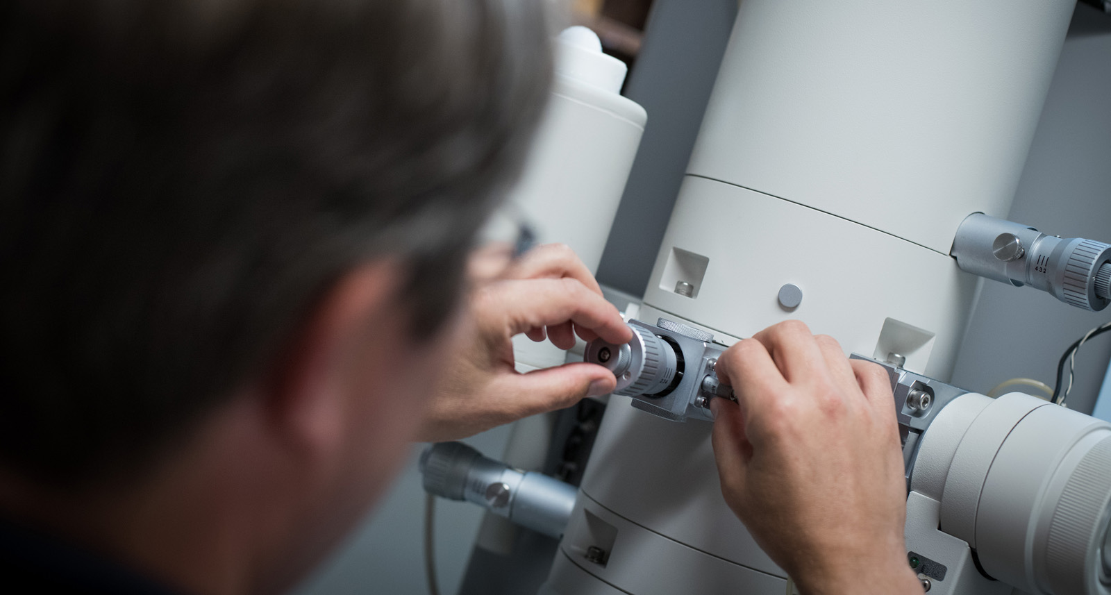
Cellulose nanocrystals are a high-strength, lightweight and resilient material extracted from trees that have a wide range of potential industrial applications. Dr. Wadood Hamad of FPInnovations partnered with UBC’s imaging lab facilities to visualize, investigate and exploit the remarkable properties of these tiny non-toxic, renewable particles. By accessing the expert assistance and advice of UBC’s lab technicians and researchers, Dr. Hamad was able to use a Transmission Electron Microscope to obtain results that show how these cellulose nanoparticles can improve the performance of nanocomposite materials in aerospace, automotive, marine and energy applications such as windmills. The Bioimaging Facility (BIF), part of UBC Imaging Labs, has a team of technical staff with specialized expertise in operating the Hitachi H7600 Transmission Electron Microscope (TEM) and a wide variety of other imaging equipment to support industry and university researchers. Dr. Wadood Hamad, Principal Scientist at FPInnovations, has worked with the BIF at various times over the past 10 years to advance research by FPInnovations on cellulose nanocrystals, a sustainable and renewable nanomaterial extracted from forest biomass. “This is a wonderful facility with very capable people and it has been fantastic in allowing us to characterize and understand these nanomaterials,” he says. In a number of projects, Dr. Hamad provided microscopy technician Brad Ross with samples of a variety of polymeric systems for TEM imaging. The Hitachi TEM has the very high spatial resolution capabilities suitable for imaging tiny nanoparticles, such as cellulose nanocrystals, that are approximately five nanometers wide and 100 nanometres long. “TEM imaging lets you see the inside of the sample, which is important because the nanocrystals are embedded in the polymer matrix,” says Brad. TEM tackles a difficult challenge The challenge in a recent project was to produce a clear image of the nanoparticles given the inherently low contrast between the polymeric systems under investigation. “It’s a very difficult challenge because the contrast between two polymer materials can be poor and difficult to distinguish between the components. If you were imaging silver or gold nanoparticles in a polymer matrix, the contrast would be much higher,” says Dr. Hamad. Brad prepared the nanocomposite sample using an ultramicrotome to cut the material into sections 50-70 nanometres thick for TEM imaging. He then experimented with various approaches to enhance the contrast between the nanocrystals and the specific polymer matrix. “The Hitachi TEM is suitable for low-contrast applications. I operated the instrument at an accelerating voltage of 80 kV because it’s easier to generate contrast at lower voltages in conjunction with the high-contrast imaging mode. I also selected a smaller objective aperture, where you sacrifice some spatial resolution for better contrast, and took advantage of the camera’s auto-gain feature, which also works to maximize contrast,” explains Brad. Imaging high-performance particles for industrial applications Dr. Hamad has used the resulting TEM images, in combination with other images and data, to build a convincing picture of these cellulose nanoparticles and their ability to improve the performance of nanocomposite materials in aerospace, automotive, marine and energy applications such as windmills. “This gives us a full suite of evidence so we can make better presentations to scientists and potential industrial collaborators, and develop applications of this material into industrial products,” he says.