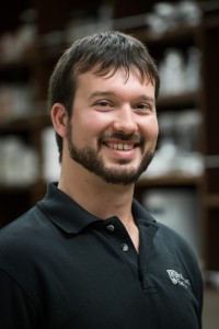
Amgen senior scientist Dr. Christopher Murawsky (Photo: Mike Dobbs)
Therapeutic monoclonal antibodies are a relatively new class of drugs used to treat patients with serious illnesses. They have shown considerable efficacy in the treatment of a variety of diseases and generally carry fewer side effects than traditional drugs. Dr. Christopher Murawsky of Amgen Inc., a leading global biotech company, partnered with UBC’s Imaging Lab facilities to visualize and analyze engineered protein nanoparticles his lab had developed to aid in the discovery of novel therapeutic antibodies. By accessing the expert assistance and advice of UBC’s lab technicians and researchers, Dr. Murawsky was able to use a Transmission Electron Microscope (TEM) to verify that these nanoparticles had the desired characteristics his team was looking for. These results contributed to Amgen’s development of a potentially useful tool for the discovery of new antibody-based therapies. Dr. Christopher Murawsky, a Senior Scientist at the Amgen research facility in Burnaby, British Columbia, worked with the Bioimaging Facility (BIF), part of UBC Imaging Labs, on a project to help develop a novel platform for the discovery of promising targets for new antibody-based therapies. Amgen is an independent global biotech company that has been a leader in developing innovative medicines including highly targeted therapies – known as monoclonal antibodies — for the treatment of serious illnesses. In collaboration with Dr. Murawsky, BIF microscopy technician Brad Ross used the Hitachi H7600 Transmission Electron Microscope (TEM) to visualize and analyze the size, shape and internal particle structure of protein-based nanoparticles that the Amgen scientists had produced in their Burnaby research lab. The tiny, self-assembling protein nanoparticles come in two distinct sizes: particles made up of 180 and 240 repeating identical subunits, ranging from 30 to 35 nanometres in diameter. Gold standard for visualizing protein nanoparticles The Amgen researchers created a modified version of the nanoparticles, in which a foreign peptide is inserted into the extended spike, allowing it to be displayed to the immune system. Dr. Murawsky’s research group is developing these modified protein nanoparticles as an alternative method for generating new monoclonal antibodies with the goal of discovering new antibody therapies. “The challenge for us was to understand this material and see if we had made intact protein nanoparticles. We wanted to determine if the modified particles were properly formed and in the right size range of 30 to 40 nanometres. TEM imaging is the gold standard for verifying and confirming that we had made the particles correctly,” explains Dr. Murawsky, Senior Scientist, Biologic Discovery. The Bioimaging Facility has a team of technical staff with specialized expertise in operating the Hitachi TEM and a wide variety of other imaging equipment to support industry and university researchers. In this project, Dr. Murawsky provided Brad with samples in solution containing the protein nanoparticles. Imaging of the samples was performed in a number of sessions over several months as the Amgen researchers developed and fine-tuned their methods of making both the control and modified nanoparticles. The Hitachi TEM has the high-contrast capabilities and high-quality imaging modes for a wide range of magnifications suitable for imaging tiny protein nanoparticles. “TEM allows you to see the internal structural details of these protein nanoparticles with much greater clarity than other methods. The Hitachi TEM is also very good at providing contrast for biological materials of this nature,” explains Brad, who applied a negative stain with 2% uranyl acetate (UA) to enhance the contrast between the background of the image and the particle samples.

BIF Technician Brad Ross

BIF research manager, Garnet Martens using the Hitachi H7600 TEM
Proof of platform with the desired features
The resulting images provided the evidence Dr. Murawsky needed to show that his research team could consistently produce protein nanoparticles with the desired features. “The TEM equipment worked very well for us. The images confirmed that we were making correctly sized and correctly shaped protein nanoparticles, and that the particles behaved properly. We now have an efficient method for making these particles because of what we learned from the images. The next step is to determine if our modified nanoparticles are useful for generating new antibodies that may be used for therapeutic or research purposes,” he says.
Getting the best images for the client’s purpose
Brad worked closely with Dr. Murawsky during the imaging process to produce useful images of the nanoparticles at different magnifications. Lower-magnification images, for example, confirmed that large groups of nanoparticles were properly formed and of the expected size. Close-up images of individual particles showed that the modified nanoparticles were distinct from but approximately the same size as control nanoparticles, and confirmed that the modified nanoparticles maintained the critical capacity to self-assemble.
“The staff are easy to work with and interested in helping the client get the images they need. Brad encouraged me to be there during the imaging so I could advise him about the kinds of images I wanted and the features I was looking for. It was a very collaborative process, which allowed us to take the best images for our purpose. The work done at the UBC Bioimaging Facility helped us immensely and contributed to us developing this platform for additional studies,” says Dr. Murawsky.