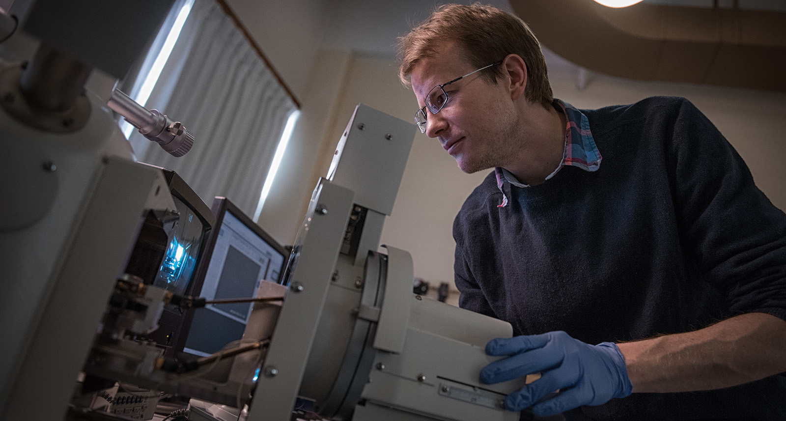
UBC Imaging Labs technician Jacob Kabel used a scanning electron microscope, equipped with an x-ray detector, to find out why a stainless steel kitchen knife had snapped without warning while he was cutting cheese. High-resolution images and analysis revealed water-induced rust as the culprit, which caused the material to slowly weaken and then completely fracture under the load of cutting. By accessing the expert training, assistance and advice of UBC’s technicians, researchers and a materials engineer, industry quality assurance staff and university researchers can use the information obtained from SEM imaging and quantitative analysis to investigate most material failures and learn how a part failed. Jacob Kabel, Electron Microscopist and Manager of the Materials Engineering Electron Microscopy Lab (MEEML), part of UBC Imaging Labs, was cutting cheese with a stainless steel knife when the blade suddenly snapped and flew past his head. Jacob wanted to know why the steel blade broke and used the facility’s Hitachi S3000N tungsten filament Scanning Electron Microscope (SEM) with Energy Dispersive X-ray (EDX) detector to investigate the cause of the material failure. “A scanning electron microscope equipped with an x-ray detector is an ideal instrument for this situation. The wide depth of field allows you to see large areas of the fracture surface in a single image and follow up with high resolution images of the microstructure. The x-ray detector can give you detailed elemental information about corrosion products, or contaminants. An SEM is a solid first choice for investigating most material failures. A materials engineer, typically working through an external consulting firm, can use the information obtained from SEM/EDX analysis to tell the story of how a part failed,” says Jacob. To prepare the sample for imaging, Jacob sawed off a segment of the stainless steel blade about a centimeter from the fracture point, so the material would fit properly into the SEM sample chamber. SEM imaging of the fracture surface provided clear visual evidence of where the crack in the knife was initiated and how far the crack propagated before it resulted in a complete fracture when the stainless steel material failed while cutting the cheese. Imaging reveals corrosion as the culprit The high-resolution images also revealed the cause of the material failure. “The knife had some corrosion that was hidden by the wooden handle and the images of the fracture surface showed corrosion where the fracture had initiated. Water had seeped in between the wooden handle of the knife and the metal. You could tell the fracture had been going on for some time and that the material was already failing because the corrosion product — the rust — could be seen clearly in the fracture. Every time the knife was used it opened up the crack a little more until the material could no longer hold under the load of cutting something,” explains Jacob. The Materials Engineering Electron Microscopy Lab has an electron microscopy technician with specialized expertise in operating the Hitachi S3000N SEM with EDX and a variety of other imaging equipment to support industry and university researchers. Jacob performed an SEM/EDX analysis of this sample after preparing a polished cross-section, which provided additional information about the concentrations of various elements such as chromium, nickel and iron in the stainless steel material. “Quantitative SEM/EDX analysis can determine the composition of various elements in a material sample to a high degree of accuracy, if done properly. That requires technical expertise and a set of standards. Our facility has SEM equipment with a full EDX analysis system and we can perform quantitative analysis with EDX rapidly and easily for a variety of industry and research applications,” says Jacob.