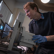
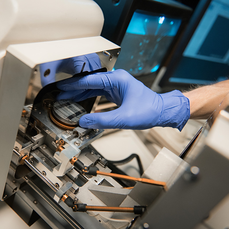
The Materials EM lab operated under the Department of Materials Engineering offers scanning electron microscopy services as well as basic xray diffraction services. By accessing the expert training, assistance and advice of UBC’s technicians, researchers and a materials engineer, industry quality assurance staff and university researchers can use the information obtained from SEM and EDX imaging and quantitative analysis to investigate materials to understand issues such as material failures and mineral composition.
Exposing root causes of material failure

UBC Imaging Labs technician Jacob Kabel used a scanning electron microscope, equipped with an x-ray detector, to find out why a stainless steel kitchen knife had snapped without warning while he was cutting cheese. High-resolution images and analysis revealed water-induced rust as the culprit, which caused the material to slowly weaken and then completely fracture under the load of cutting.
Assessing composition and compatibility of ore grinding balls

Imasco Minerals had an opportunity to buy several hundred steel balls used to crush and grind ore in a ball mill. But the company needed to have accurate and detailed information about the composition of the steel balls to assess whether these would be compatible with its existing ball milling equipment. Imasco quality assurance manager David Quan worked with UBC’s imaging lab facilities to analyze the concentrations of iron, carbon, chromium, manganese and other elements in three sizes of steel balls. The imaging and analysis revealed some important similarities and unexpected differences in the composition of the small balls, compared with the medium and large balls. By accessing the expert assistance and advice of UBC’s lab technicians and researchers, Mr. Quan was able to use a Hitachi scanning electron microscope, equipped with an x-ray detector, to learn that the composition of the various steel balls was similar enough to the ones Imasco currently uses to consider buying them.