
New ways to tackle some of the biggest health issues we are facing are being developed by researchers utilizing the micro-CT capabilities of UBC’s Centre for High-Throughput Phenogenomics (CHTP). Taking advantage of CHTP’s highest quality x-ray imaging equipment combined with extensive technical support, pre-approved research protocols and access to animal care facilities, researchers are developing new tools to study the progression of chronic obstructive pulmonary disease (COPD) and are investigating the link between obesity and insulin levels.
Longitudinal studies with non-invasive in vivo imaging
A key benefit of utilizing the Trifoil eXplore CT 120 in vivo scanner is that it is non-invasive in nature, allowing researchers to monitor changes over time in the same subjects in small animal model studies. This not only provides a better way to follow disease progression and monitor the response to treatments, it also significantly reduces the number of animals required for any research program.
Gating Techniques
Dr. Nancy Ford
Dr. Nancy Ford, CHTP lab Director and a UBC medical physics researcher, is using in vivo scanning to develop new imaging techniques to investigate anatomical and functional changes over time in COPD, a progressive lung disease that affects more than 200 million people worldwide, and is projected to be the third leading cause of death by 2030. Currently, the disease process is normally assessed using histology, which lacks sensitivity to detect subtle but important changes in the lungs, which occur early in human COPD. Dr. Ford’s micro-CT tool could provide a more natural and realistic way for researchers to model and study lung structure and function in early COPD and then follow disease progression.
In developing her imaging tool, Dr. Ford uses a micro-CT respiratory gating technique to obtain sharper, more accurate images of airways, blood vessels and other small structures within the lungs of rodents than could be achieved with non-gated imaging. The technique synchronizes the image data with the rodent’s respiratory signal to reconstruct 3D images at prescribed phases, such as full inspiration and expiration. The images acquired through respiratory gating can be analyzed quantitatively to characterize anatomical and functional changes in rodent models of COPD and other respiratory diseases. “In a clinic, you can tell humans to hold their breath, but you can’t do that with free-breathing animals. Rodents breathe very quickly and their lungs move a lot during the imaging session. Respiratory gating reduces the motion artifacts and blurring of the images caused by their movements,” she says.
Dr. Ford also applied the dual-energy imaging capabilities of the scanner, along with iodine and xenon contrast agents, to selectively enhance, contrast and differentiate blood vessels from soft tissues.” The contrast agents are important for measuring differences in lung function as well, such as air content in the lungs after exhalation. “Areas with reduced concentrations of the contrast agents appear less bright and suggest potential airflow obstructions in different regions of the lungs,” she explains.
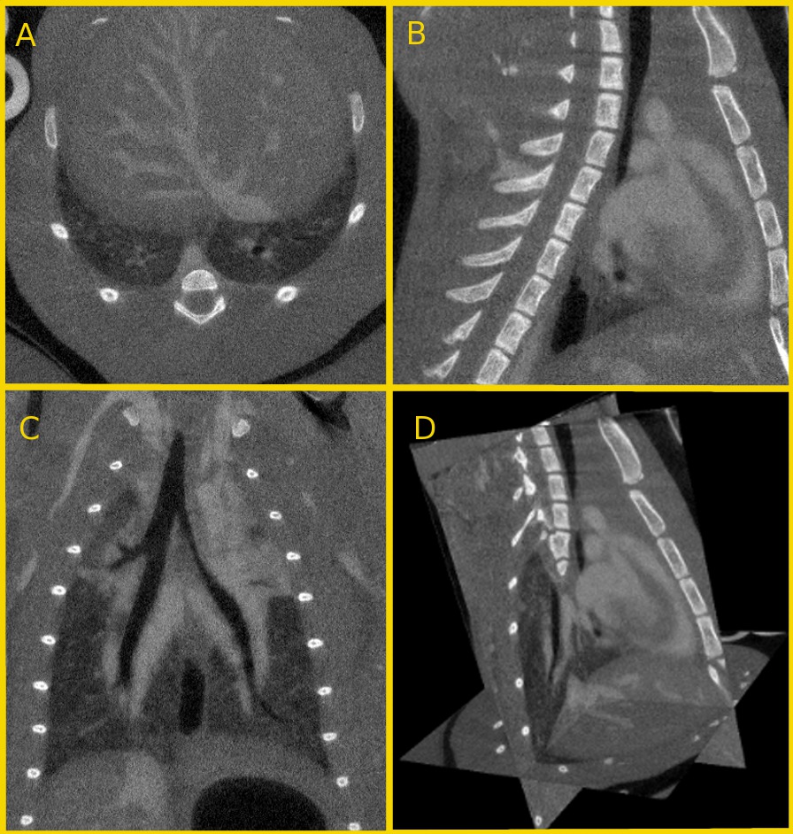
Contrast-enhanced micro-CT images of a mouse thorax and upper abdomen. A) Transverse section of the liver vasculature and inferior portion of the lungs; B) Sagittal section of the heart between the spine (left) and sternum (right); C) Coronal section of the lungs; D) 3D dataset automatically reconstructed from the micro-CT scan.
Specimen scanning
To complement the in vivo results, Dr. Ford uses the Scanco Medical µCT100 micro-CT specimen scanner at the CHTP to investigate lung tissues. “The specimen scanner allows researchers to image the lungs at a higher resolution to see smaller blood vessels and it has enabled us to get useful and more detailed quantitative information about different anatomical structures and regions in the lungs,” she says.
Support for new users
Researchers in the lab of Dr. James Johnson utilized CHTP’s micro-CT capabilities to examine the role of elevated insulin levels (hyperinsulinemia) in obesity, a health condition that affects more than 500 million people worldwide. A key element in the success of their project was the technical and research support Dr. Skovsø, a postdoctoral researcher in the lab, received from CHTP staff.
Dr. Skovsø worked closely with John Schipilow, one of CHTP’s skilled technicians, on both the imaging and the data analysis. John performed the initial CT scans of the control and experimental mice with Dr. Skovsø present during the imaging sessions and provided guidance on using the software to perform the data analysis. His expertise, combined with Dr. Skovsø’s background knowledge on rodent fat distribution imaging, enabled her to calculate the volumes for the fat tissue regions she wanted to measure. “John had a great deal of knowledge about the machine and CT scanning in general. He trained me so that gradually over time I started doing more of the scanning and was able to do the data analysis on my own. He was very patient and helped me to overcome the challenges I needed to solve,” says Dr. Skovsø.
The project was also able to take advantage of using a pre-approved protocol for micro-CT scanning of soft tissues like fat and muscle. By accessing this protocol, Dr. Skovsø’s imaging study was both streamlined and more effective. “Following this protocol, the micro-CT scanner generates images of good quality with a low radiation dose. Reducing radiation dose allows for multiple scan acquisitions over time to follow these animals,” she explains.
Having simple access to animal care facilities is a further advantage. As John explains, “A big advantage of doing micro-CT at this facility is that researchers can get easy access to their animals, perform the imaging, and do their analysis in a sequential manner in the same place with as much training and support as they need from expert technical staff. The researcher can focus on his or her study, without having to worry about the technical aspects.”

Left: A cross sectional in vivo micro-CT image of a murine visceral fat compartment and subcutaneous fat compartment
Right: Visceral fat (red) and subcutaneous fat (blue) modelled in 3D allows for the visual representation and volumetric quantification of each respective compartment.
Influential results and ongoing partnerships
These micro-CT capabilities have had a huge impact on both research studies and are paving the way for ongoing collaboration and partnerships.
The tool being developed by Ford could provide a more natural and realistic way for researchers to model and study lung structure and function. “With this imaging tool,” says Ford, “we hope to see symptoms like airflow obstructions at the early stage, and be able to accurately measure changes in the blood vessels, airways and other lung system components in pre-clinical models of COPD. We’ve done the preliminary experiments in healthy rodents that show we can image the same animal a number of times and we’re able to quantify what we see in the images. We’re now optimizing the technique to make sure the measurements of lung function and structures are accurate. The next step will be to test it in animal models of COPD,” she says.
For Dr. Skovsø and researchers in Dr. Johnson’s lab, utilizing micro-CT imaging at CHTP provided greater accuracy and specificity compared to other methods such as micro-DEXA. It also allowed them to look at changes in fat distribution in the same mice over time. This enabled them to accurately visualize and measure changes in both the harmful and less harmful types of abdominal fat that resulted from lowering insulin levels and show that lowering insulin production to normal levels reverses weight gain in adult mice fed a high-fat diet.
These promising results suggest that reducing high insulin levels through diet or drugs could potentially help people to lose weight and reduce the harmful type of fat associated with type 2 diabetes.
Having learned how to use micro-CT scanning to analyze fat tissues, and knowing its capabilities, this is just the start of a longer relationship between Dr. Skovsø, her colleagues and CHTP. “The researchers in our lab, including myself, will likely use micro-CT a lot to look at fat distribution in other obesity and type 2 diabetes studies,” she says.
In doing so they will be joining a wide range of research partners that are taking advantage of CHTP’s micro-CT capacity.